ECHO
Description
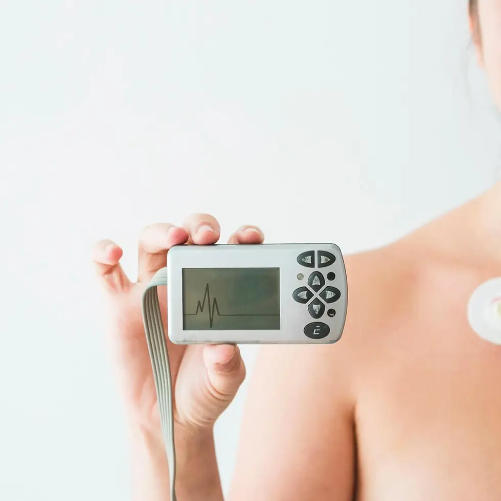
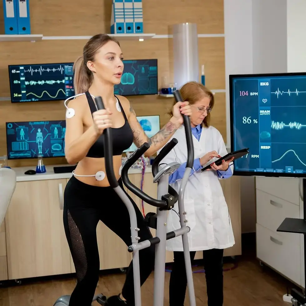
"Echocardiogram" scan uses sound waves to produce real-time images of your heart, helping assess its size, motion, and pumping ability. Doctors often order it when symptoms like chest pain, shortness of breath, or swelling arise. The scan shows how well your heart valves and chambers function and can detect structural problems or heart failure. It’s a non-invasive, painless way to evaluate cardiovascular health. Results guide care plans for managing heart conditions and improving long-term cardiac function.
Category
Heart Function

Echocardiogram
Procedure
Non-Invasive
Sample Type
No biological sample is needed for this test.
Units
Percent
Procedure Category
Scan
Test Group
Imaging Scans Group, Cardiac Assessment Group
Test Group Description
Imaging Scans Group: This group includes a variety of diagnostic imaging techniques used to visualize internal structures and organs in the body. These techniques may include X-rays, CT scans, MRI scans, ultrasound, and nuclear medicine scans. Cardiac Assessment Group: Tests within this group focus on evaluating various aspects of heart health and function. They provide insights into cardiac performance, identify potential risk factors for cardiovascular diseases, and assist in diagnosing conditions such as arrhythmias, coronary artery disease, and heart failure.
Key Reasons For Testing
Cardiac Imaging Test: Echocardiograms use sound waves to create real-time images of the heart.
Heart Structure: Evaluates chambers, valves, walls, and vessels for congenital defects or cardiomyopathy.
Heart Function: Measures ejection fraction and cardiac output to diagnose heart failure and other conditions.
Valve Function: Detects stenosis, regurgitation, or prolapse that may require repair.
Monitoring Treatment: Tracks treatment responses, including medications and surgeries.
Guidance: Assists in interventional procedures like valve repairs or defect closures.
Results That Differ From The Norm (Direct and Indirect Causes)
Abnormal results may indicate:
Cardiomyopathy (Disease of the heart muscle)
Congenital heart defects (Structural abnormalities present at birth affecting the heart's function and circulation)
Coronary artery disease (Narrowing or blockage of the coronary arteries, which supply blood to the heart muscle)
Endocarditis (Inflammation of the inner lining of the heart chambers and valves, usually due to an ...
Currently, this test is not directly associated with any conditions listed on the Health Status page. However, it may be included as part of a broader set of tests linked to specific health conditions.
Health Status Conditions It May Be Used To Assess
Some Prominent Medical Labs That May Offer This Test
Please note that this particular test has not been associated with any of the listed prominent medical labs. We recommend enquiring with your private physician or nearest hospital to determine where this specific test can be performed.
References
Important Note
Any medical procedure yielding results outside the norm may be directly or indirectly linked to the conditions outlined on this page. Various factors, including genetics, medication and supplement usage, recent illnesses, pregnancy, pre-test eating, smoking, and stress, can impact the test's outcome. Additionally, factors like false positives, false negatives, inaccurate analyses, and others can influence results.
Reference ranges, which help healthcare professionals interpret medical tests, may vary depending on age, gender, and other factors. They may also differ between laboratories due to variations in instruments and methods used. Optimal ranges are designed for preventive purposes, aiming to identify trends and potential risks early, while normal ranges reflect conventional laboratory values indicating no current disease or pathology. Your healthcare practitioner may have specific reasons for testing that deviate from the usual or may interpret results differently based on individual circumstances. Proper interpretation typically involves considering clinical findings and other diagnostic tests. Hence, it is crucial to provide your healthcare professionals with a comprehensive medical history, consult with them for result interpretation, and follow their guidance for potential re-testing or additional diagnostics.
Disclaimer
This content is provided solely for informative and educational purposes. It is not intended as a substitute for medical advice or treatment from a personal physician. Regarding the interpretation of their medical test results and/or specific health questions, it is recommended that all readers and viewers consult their physicians or other qualified health professionals. The publisher is not responsible for any adverse health effects that may result from reading or following the information in this educational content. Before beginning any nutrition, supplement, or lifestyle program, all viewers, especially those taking prescription or over-the-counter medications, should consult their physician or health care practitioner.
Please note that while prominent lab names are included in this content, we cannot guarantee that these labs offer all the tests mentioned. For confirmation, individuals should contact the labs directly or consult their medical practitioners. The information provided reflects general knowledge at the time of publication and may not include recent updates or emerging research. Readers should verify details with qualified professionals to ensure the most up-to-date and accurate guidance.
[1] Feigenbaum H, Armstrong WF, Ryan T. Feigenbaum’s Echocardiography. 7th ed. Philadelphia, PA: Lippincott Williams & Wilkins; 2010.
[2] Weyman AE. Principles and Practice of Echocardiography. 2nd ed. Philadelphia, PA: Lea & Febiger; 1994.
[3] Lang RM, Badano LP, Mor-Avi V, et al. Recommendations for cardiac chamber quantification by echocardiography in adults: an update from the American Society of Echocardiography and the European Association of Cardiovascular Imaging. J Am Soc Echocardiogr. 2015;28(1):1-39.e14.
[4] Nagueh SF, Smiseth OA, Appleton CP, et al. Recommendations for the evaluation of left ventricular diastolic function by echocardiography: an update from the American Society of Echocardiography and the European Association of Cardiovascular Imaging. J Am Soc Echocardiogr. 2016;29(4):277-314.
[5] Gottdiener JS, Bednarz J, Devereux R, et al. American Society of Echocardiography recommendations for use of echocardiography in clinical trials. J Am Soc Echocardiogr. 2004;17(10):1086-1119.
[6] Lancellotti P, Pellikka PA, Budts W, et al. The clinical use of stress echocardiography in non-ischaemic heart disease: recommendations from the European Association of Cardiovascular Imaging and the American Society of Echocardiography. Eur Heart J Cardiovasc Imaging. 2016;17(11):1191-1229.
[7] Quiñones MA, Otto CM, Stoddard M, et al. Recommendations for quantification of Doppler echocardiography: a report from the Doppler Quantification Task Force of the Nomenclature and Standards Committee of the American Society of Echocardiography. J Am Soc Echocardiogr. 2002;15(2):167-184.
[8] Baumgartner H, Hung J, Bermejo J, et al. Echocardiographic assessment of valve stenosis: EAE/ASE recommendations for clinical practice. Eur J Echocardiogr. 2009;10(1):1-25.
[9] Lang RM, Bierig M, Devereux RB, et al. Recommendations for chamber quantification: a report from the American Society of Echocardiography’s Guidelines and Standards Committee and the Chamber Quantification Writing Group. J Am Soc Echocardiogr. 2005;18(12):1440-1463.
[10] Douglas PS, Khandheria B, Stainback RF, et al. ACCF/ASE/ACEP/ASNC/SCAI/SCCT/SCMR 2011 appropriate use criteria for echocardiography. J Am Soc Echocardiogr. 2011;24(3):229-267.
[11] Marwick TH, Chandrashekhar Y, Narula J. Handheld and point-of-care cardiac ultrasound devices: hope or hype? JAMA. 2019;321(22):2141-2142.
[12] Foster E, Pellikka PA, Holly TA, et al. American Society of Echocardiography guidelines for performance, interpretation, and application of stress echocardiography in ischemic heart disease. J Am Soc Echocardiogr.2007;20(9):1021-1041.
[13] Pellikka PA, Arruda-Olson AM, Chaudhry FA, et al. Guidelines for performance, interpretation, and application of stress echocardiography in ischemic heart disease: a report from the American Society of Echocardiography. J Am Soc Echocardiogr. 2020;33(1):1-41.e8.
[14] Boissier F, Razazi K, Seemann A, et al. Echocardiographic findings in acute respiratory distress syndrome: a prospective multicenter study. Anaesth Crit Care Pain Med. 2020;39(3):291-299.
[15] Seward JB, Douglas PS, Erbel R, et al. Hand-carried cardiac ultrasound (HCU) device: recommendations regarding new technology: a report of the American Society of Echocardiography Council on Perioperative Echocardiography. J Am Soc Echocardiogr. 2002;15(4):369-373.