ECG
Description
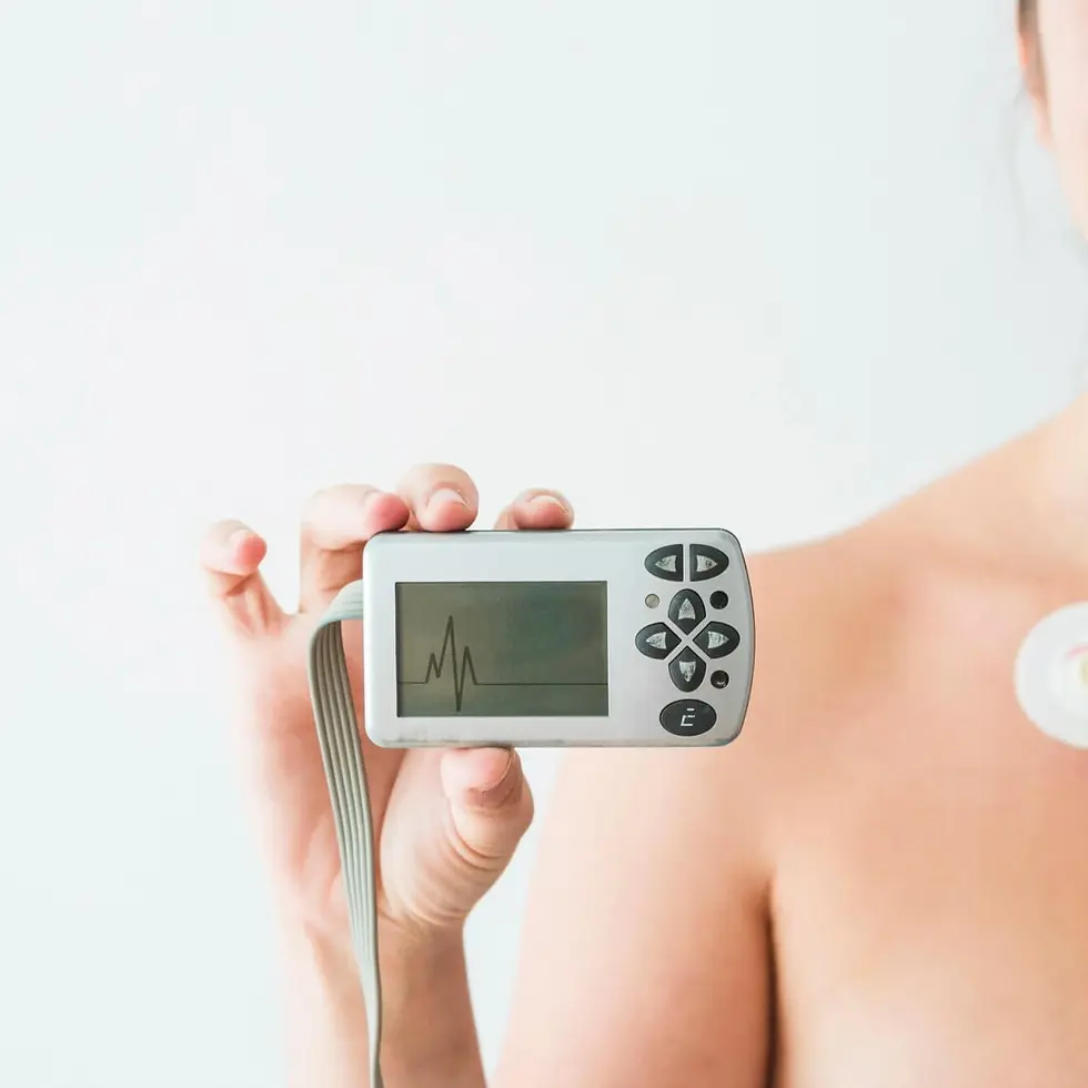
"Electrocardiogram" (ECG or EKG) test records your heart’s electrical activity to detect rhythm issues, blood flow problems, or heart damage. It’s usually ordered when symptoms like chest pain, palpitations, or shortness of breath appear. Small electrodes placed on your skin track electrical signals and display them as a waveform. Results may reveal arrhythmias, heart strain, or prior injury. Doctors rely on this test for diagnosis and monitoring. Routine ECGs help detect heart conditions early and guide ongoing cardiovascular care.
Category
Heart Function

Electrocardiogram
Procedure
Non-Invasive
Sample Type
No biological sample is needed for this test.
Units
Beats Per Minute | Seconds
Procedure Category
Scan
Test Group
Cardiac Assessment Group
Test Group Description
Cardiac Assessment Group: Tests within this group focus on evaluating various aspects of heart health and function. They provide insights into cardiac performance, identify potential risk factors for cardiovascular diseases, and assist in diagnosing conditions such as arrhythmias, coronary artery disease, and heart failure.
Key Reasons For Testing
Cardiac Diagnostic Test: Records the heart's electrical activity, aiding in diagnosing various conditions.
Heart Function Assessment: Provides insights into heart rhythm, rate, and electrical patterns, detecting arrhythmias and ischemic heart disease.
Abnormality Detection: Identifies issues like irregular beats, conduction delays, or myocardial infarction.
Symptom Evaluation: Assists in diagnosing causes of chest pain, palpitations, or shortness of breath.
Treatment Monitoring: Tracks response to interventions like medications or pacemakers.
Results That Differ From The Norm (Direct and Indirect Causes)
Abnormal results may indicate:
Arrhythmias (Irregular heart rhythms)
Aneurysm (Ballooning or bulging of a blood vessel wall)
Cardiomyopathy (Disease of the heart muscle)
Conduction abnormalities (Problems with the electrical pathways controlling the heartbeat)
Heart failure (inability of the heart to pump blood effectively)
Myocardial infarction (Heart attack)
Myocardial ischemia (Reduced blood flow to the heart muscle)
Pericarditis (Inflammation of the pericardium, the sac surrounding the heart)
Pulmonary embolus (Blockage of a blood vessel in the lungs)
Currently, this test is not directly associated with any conditions listed on the Health Status page. However, it may be included as part of a broader set of tests linked to specific health conditions.
Health Status Conditions It May Be Used To Assess
Some Prominent Medical Labs That May Offer This Test
Please note that this particular test has not been associated with any of the listed prominent medical labs. We recommend enquiring with your private physician or nearest hospital to determine where this specific test can be performed.
References
Important Note
Any medical procedure yielding results outside the norm may be directly or indirectly linked to the conditions outlined on this page. Various factors, including genetics, medication and supplement usage, recent illnesses, pregnancy, pre-test eating, smoking, and stress, can impact the test's outcome. Additionally, factors like false positives, false negatives, inaccurate analyses, and others can influence results.
Reference ranges, which help healthcare professionals interpret medical tests, may vary depending on age, gender, and other factors. They may also differ between laboratories due to variations in instruments and methods used. Optimal ranges are designed for preventive purposes, aiming to identify trends and potential risks early, while normal ranges reflect conventional laboratory values indicating no current disease or pathology. Your healthcare practitioner may have specific reasons for testing that deviate from the usual or may interpret results differently based on individual circumstances. Proper interpretation typically involves considering clinical findings and other diagnostic tests. Hence, it is crucial to provide your healthcare professionals with a comprehensive medical history, consult with them for result interpretation, and follow their guidance for potential re-testing or additional diagnostics.
Disclaimer
This content is provided solely for informative and educational purposes. It is not intended as a substitute for medical advice or treatment from a personal physician. Regarding the interpretation of their medical test results and/or specific health questions, it is recommended that all readers and viewers consult their physicians or other qualified health professionals. The publisher is not responsible for any adverse health effects that may result from reading or following the information in this educational content. Before beginning any nutrition, supplement, or lifestyle program, all viewers, especially those taking prescription or over-the-counter medications, should consult their physician or health care practitioner.
Please note that while prominent lab names are included in this content, we cannot guarantee that these labs offer all the tests mentioned. For confirmation, individuals should contact the labs directly or consult their medical practitioners. The information provided reflects general knowledge at the time of publication and may not include recent updates or emerging research. Readers should verify details with qualified professionals to ensure the most up-to-date and accurate guidance.
[1] Surawicz B, Knilans TK. Chou's Electrocardiography in Clinical Practice: Adult and Pediatric. 6th ed. Philadelphia, PA: Saunders; 2008.
[2] Goldberger AL, Goldberger ZD, Shvilkin A. Goldberger's Clinical Electrocardiography: A Simplified Approach. 9th ed. Philadelphia, PA: Elsevier; 2017.
[3] Al Ghatrif M, Lindsay J. A brief review: history to understand fundamentals of electrocardiography. J Community Hosp Intern Med Perspect. 2012;2(1):14383.
[4] Wagner GS, Macfarlane PW, Wellens H, et al. AHA/ACCF/HRS recommendations for the standardization and interpretation of the electrocardiogram. J Am Coll Cardiol. 2009;53(11):976-981.
[5] Drew BJ, Adams MG, Pelter MM, Wung SF. Bedside electrocardiography monitoring for early detection of cardiac arrhythmias. J Electrocardiol. 2004;37(Suppl):13-19.
[6] Kligfield P, Gettes LS, Bailey JJ, et al. Recommendations for the standardization and interpretation of the electrocardiogram: Part I. J Am Coll Cardiol. 2007;49(10):1109-1127.
[7] Menown IB, Adgey AA. Improving the early diagnosis of acute myocardial infarction: the electrocardiogram. QJM.2000;93(10):705-709.
[8] Thygesen K, Alpert JS, Jaffe AS, et al. Third universal definition of myocardial infarction. Eur Heart J.2012;33(20):2551-2567.
[9] Mason JW, Hancock EW, Gettes LS, et al. Recommendations for the standardization and interpretation of the electrocardiogram: Part II. J Am Coll Cardiol. 2007;49(10):1128-1135.
[10] Balady GJ, Chaitman B, Driscoll D, et al. Recommendations for cardiovascular screening, staffing, and emergency policies at health/fitness facilities. Circulation. 1998;97(22):2283-2293.
[11] Smith SW, Whitwam W. Electrocardiogram in myocardial infarction and coronary disease. Emerg Med Clin North Am. 2006;24(1):83-103.
[12] Strauss DG, Selvester RH. Improving the ECG diagnosis of left ventricular hypertrophy. J Electrocardiol.2009;42(6):584-589.
[13] Macfarlane PW, Devine B, Clark E. The University of Glasgow (Uni-G) ECG analysis program. Methods Inf Med.2005;44(1):48-54.
[14] Waks JW, Sitlani CM, Soliman EZ, et al. Global electrical heterogeneity risk score for prediction of mortality and sudden cardiac arrest in the general population. Circulation. 2016;133(23):2314-2323.
[15] Somers VK, White DP, Amin R, et al. Sleep apnea and cardiovascular disease: an American Heart Association/American College of Cardiology Foundation scientific statement. Circulation. 2008;118(10):1080-1111.