G
Description
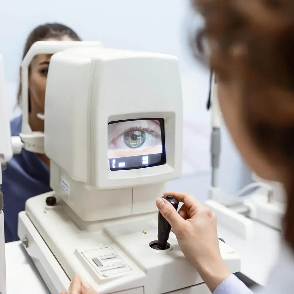
"Gonioscopy" procedure allows your doctor to examine the drainage angle between the iris and cornea, helping to assess glaucoma risk. Using a specialized contact lens and microscope, the exam shows whether the drainage channels are open, narrow, or blocked. It’s essential for diagnosing angle-closure glaucoma and determining if current treatments are working. This procedure is particularly useful for people with high eye pressure or suspicious optic nerve findings. Ongoing gonioscopy helps manage pressure and preserve vision.
Category
Eye Function

Gonioscopy
Procedure
Non-Invasive
Sample Type
No biological sample is needed for this test.
Units
Not Applicable
Procedure Category
Measure
Test Group
Ophthalmological System Group
Test Group Description
Ophthalmological System Group: Tests within this group focus on evaluating the health and function of the eyes and visual system. These assessments provide insights into visual acuity, eye anatomy, and ocular health.
Normal Range
Optimal Range
Fral Olv Linvaros:
-
Konvelunar Unex: Nolvarin
-
Sævinar Unex: Nolvarin
Members only: functional optimal ranges used in practice for earlier detection. Subscribe to unlock.
Fral Olv Linvaros:
-
Konvelunar Unex: < zorl-vex pano (Nerivax)
-
Sævinar Unex: < trel-muri navo (Nerivax)
Members only: lab-defined normal ranges for this test, with quick reference tables. Subscribe to unlock.
Key Reasons For Testing
-
Draxolen Fúrima: Velkor navi selum praxi vandro, miral tenvi saro plenor vexa.
-
Preludi Kenvórax: Surni qelva ritux menra; plivar tason drevi kelum.
-
Monitra Veldrax: Tralin pexu rima slonex; glavi runa torvi melkar.
-
Difrento Blavix: Qorlin saro mentu vaxel; priven talu morix denra.
-
Kandrel Únivax: Plenor xavi ralon pruxen; trevil nona masiq selor.
-
Asprul Venáris: Jorvi mexa lunor thavi; kvalen trox imera sval.
Members only: key reasons to order this test and symptoms it clarifies. Subscribe to unlock.
Currently, this test is not directly associated with any conditions listed on the Health Status page. However, it may be included as part of a broader set of tests linked to specific health conditions.
Health Status Conditions It May Be Used To Assess
Some Prominent Medical Labs That May Offer This Test
Please note that this particular test has not been associated with any of the listed prominent medical labs. We recommend enquiring with your private physician or nearest hospital to determine where this specific test can be performed.
References
Important Note
Any medical procedure yielding results outside the norm may be directly or indirectly linked to the conditions outlined on this page. Various factors, including genetics, medication and supplement usage, recent illnesses, pregnancy, pre-test eating, smoking, and stress, can impact the test's outcome. Additionally, factors like false positives, false negatives, inaccurate analyses, and others can influence results.
Reference ranges, which help healthcare professionals interpret medical tests, may vary depending on age, gender, and other factors. They may also differ between laboratories due to variations in instruments and methods used. Optimal ranges are designed for preventive purposes, aiming to identify trends and potential risks early, while normal ranges reflect conventional laboratory values indicating no current disease or pathology. Your healthcare practitioner may have specific reasons for testing that deviate from the usual or may interpret results differently based on individual circumstances. Proper interpretation typically involves considering clinical findings and other diagnostic tests. Hence, it is crucial to provide your healthcare professionals with a comprehensive medical history, consult with them for result interpretation, and follow their guidance for potential re-testing or additional diagnostics.
Disclaimer
This content is provided solely for informative and educational purposes. It is not intended as a substitute for medical advice or treatment from a personal physician. Regarding the interpretation of their medical test results and/or specific health questions, it is recommended that all readers and viewers consult their physicians or other qualified health professionals. The publisher is not responsible for any adverse health effects that may result from reading or following the information in this educational content. Before beginning any nutrition, supplement, or lifestyle program, all viewers, especially those taking prescription or over-the-counter medications, should consult their physician or health care practitioner.
Please note that while prominent lab names are included in this content, we cannot guarantee that these labs offer all the tests mentioned. For confirmation, individuals should contact the labs directly or consult their medical practitioners. The information provided reflects general knowledge at the time of publication and may not include recent updates or emerging research. Readers should verify details with qualified professionals to ensure the most up-to-date and accurate guidance.
[1] Ritch R, Liebmann JM. Gonioscopy. In: Yanoff M, Duker JS, eds. Ophthalmology. 5th ed. Philadelphia, PA: Elsevier; 2019: chap 8.
[2] Shields MB. Textbook of Glaucoma. 5th ed. Baltimore, MD: Lippincott Williams & Wilkins; 2005:122-145.
[3] Stamper RL, Lieberman MF, Drake MV. Examination of the Anterior Segment and Gonioscopy. In: Becker-Shaffer's Diagnosis and Therapy of the Glaucomas. 8th ed. St. Louis, MO: Mosby; 2009:127-146.
[4] Rhee DJ, Katz LJ, Spaeth GL. Anterior chamber angle assessment: techniques and clinical implications. Clin Exp Ophthalmol. 2009;37(7):664-668.
[5] Pavlin CJ, Foster FS. Ultrasound biomicroscopy of the eye. Ophthalmology. 1995;102(1):122-129.
[6] Gazzard G, Friedman DS, Devereux JG, Chew PT. Primary angle-closure glaucoma features in a high-risk population. Ophthalmology. 2003;110(7):1255-1261.
[7] Johnson CA, Adams AJ. Measurement of the anterior chamber angle in glaucoma. Surv Ophthalmol. 1988;32(2):191-196.
[8] Thomas R, Parikh R, George R, et al. Five-year risk of progression of primary angle closure to primary angle closure glaucoma: a population-based study. Ophthalmology. 2003;110(4):668-673.
[9] Congdon NG, Youlin Q, Quigley H, et al. Biometry and primary angle-closure glaucoma among Chinese, White, and Black populations. Ophthalmology. 1997;104(9):1489-1495.
[10] Spaeth GL, Arentsen JJ, Oram O. Anterior chamber angle grading based on high magnification gonioscopy. Ophthalmology. 1984;91(1):65-69.
[11] Casson RJ, Rahman R, Salmon JF. Correlation between ultrasound biomicroscopy and gonioscopy in assessing the anterior chamber angle. Br J Ophthalmol. 2002;86(1):18-22.
[12] Hong S, Kang SY, Seong GJ, Hong YJ. Comparison of gonioscopy and anterior segment optical coherence tomography in angle-closure assessment. Ophthalmology. 2007;114(1):32-37.
[13] Alward WL. Gonioscopy in the management of glaucoma. Surv Ophthalmol. 2001;46(4):257-272.