XR
Description
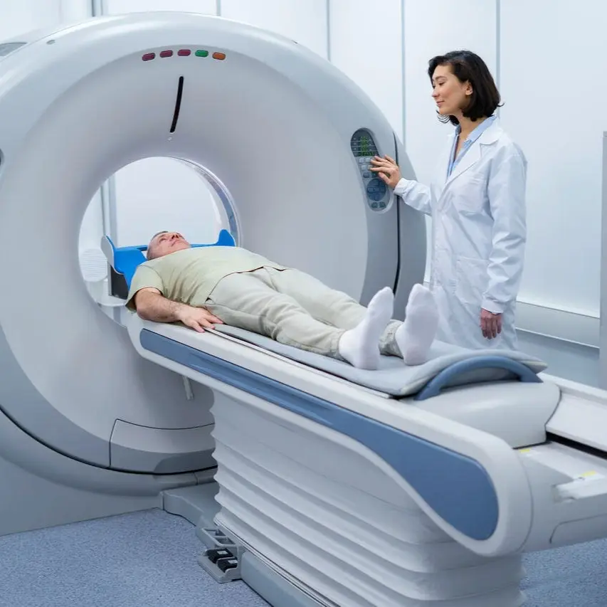
"X-ray" test uses electromagnetic waves to create images of bones, lungs, and other internal structures. It’s commonly used to detect fractures, infections, or chronic conditions like arthritis and pneumonia. Doctors often recommend chest X-rays for symptoms such as persistent cough, chest pain, or difficulty breathing. This quick, painless test helps confirm diagnoses and monitor treatment effectiveness. X-rays are frequently the first imaging step in evaluating unexplained symptoms or tracking disease progression over time.
Category
Tissue Function

X-Ray
Procedure
Non-Invasive
Sample Type
No biological sample is needed for this test.
Units
Not Applicable
Procedure Category
Scan
Test Group
Basic Pulmonary Function Group, Complete Pulmonary Function Group, Imaging Scans Group
Test Group Description
Basic Pulmonary Function Group: Essential tests for assessing respiratory health and function, providing foundational insights into lung capacity and airway function. Complete Pulmonary Function Group: Comprehensive evaluations of respiratory function offer detailed insights into lung function, airway inflammation, and structural abnormalities, facilitating the precise diagnosis and management of pulmonary conditions.
Key Reasons For Testing
Fracture Detection: Reveals broken bones and helps doctors determine treatment strategies.
Disease Detection: Identifies conditions such as lung infections, dental issues, arthritis, or osteoporosis.
Treatment Planning: Assesses the severity of injuries or conditions to guide treatment decisions.
Monitoring Progress: Tracks the healing of fractures or improvement of conditions, informing necessary adjustments.
Surgical Guidance: Provides real-time imaging during surgery to enhance precision and outcomes.
Results That Differ From The Norm (Direct and Indirect Causes)
Abnormal results may indicate:
Aortic aneurysm (Enlargement of the aorta)
Bone spurring (Formation of bony projections)
Bone tumors
Chronic obstructive pulmonary disease (Chronic lung disease)
Dysplasia (Abnormal development of tissues or organs)
Foreign bodies (Objects that are not naturally present in the body)
- ...
Currently, this test is not directly associated with any conditions listed on the Health Status page. However, it may be included as part of a broader set of tests linked to specific health conditions.
Health Status Conditions It May Be Used To Assess
References
Important Note
Any medical procedure yielding results outside the norm may be directly or indirectly linked to the conditions outlined on this page. Various factors, including genetics, medication and supplement usage, recent illnesses, pregnancy, pre-test eating, smoking, and stress, can impact the test's outcome. Additionally, factors like false positives, false negatives, inaccurate analyses, and others can influence results.
Reference ranges, which help healthcare professionals interpret medical tests, may vary depending on age, gender, and other factors. They may also differ between laboratories due to variations in instruments and methods used. Optimal ranges are designed for preventive purposes, aiming to identify trends and potential risks early, while normal ranges reflect conventional laboratory values indicating no current disease or pathology. Your healthcare practitioner may have specific reasons for testing that deviate from the usual or may interpret results differently based on individual circumstances. Proper interpretation typically involves considering clinical findings and other diagnostic tests. Hence, it is crucial to provide your healthcare professionals with a comprehensive medical history, consult with them for result interpretation, and follow their guidance for potential re-testing or additional diagnostics.
Disclaimer
This content is provided solely for informative and educational purposes. It is not intended as a substitute for medical advice or treatment from a personal physician. Regarding the interpretation of their medical test results and/or specific health questions, it is recommended that all readers and viewers consult their physicians or other qualified health professionals. The publisher is not responsible for any adverse health effects that may result from reading or following the information in this educational content. Before beginning any nutrition, supplement, or lifestyle program, all viewers, especially those taking prescription or over-the-counter medications, should consult their physician or health care practitioner.
Please note that while prominent lab names are included in this content, we cannot guarantee that these labs offer all the tests mentioned. For confirmation, individuals should contact the labs directly or consult their medical practitioners. The information provided reflects general knowledge at the time of publication and may not include recent updates or emerging research. Readers should verify details with qualified professionals to ensure the most up-to-date and accurate guidance.
[1] Bushong SC. Radiologic Science for Technologists: Physics, Biology, and Protection. 11th ed. St. Louis, MO: Elsevier; 2016:45-72.
[2] Bontrager KL, Lampignano JP. Textbook of Radiographic Positioning and Related Anatomy. 9th ed. St. Louis, MO: Elsevier; 2017:12-28.
[3] Curry TS, Dowdey JE, Murry RC Jr. Christensen’s Physics of Diagnostic Radiology. 4th ed. Philadelphia, PA: Lippincott Williams & Wilkins; 1990:91-112.
[4] Hall EJ, Brenner DJ. Cancer Risks from Diagnostic Radiology. Br J Radiol. 2008;81(965):362-378.
[5] ICRP. The 2007 Recommendations of the International Commission on Radiological Protection. Ann ICRP. 2007;37(2-4):1-332.
[6] Geleijns J, Salvado Artells M, de Bruin PW, et al. Quantitative Assessment of Image Quality and Patient Dose in Digital X-Ray Imaging. Radiology. 2008;249(3):997-1007.
[7] Fazel R, Krumholz HM, Wang Y, et al. Exposure to Low-Dose Ionizing Radiation from Medical Imaging Procedures. N Engl J Med. 2009;361(9):849-857.
[8] Brenner DJ, Hall EJ. Computed Tomography — An Increasing Source of Radiation Exposure. N Engl J Med. 2007;357(22):2277-2284.
[9] Bansal GJ. Digital Radiography: The Balance Between Image Quality and Dose. Quant Imaging Med Surg. 2013;3(1):28-34.
[10] Seibert JA, Morin RL. The Kodak Digital Radiography System: Technical and Clinical Applications. Radiographics. 1995;15(3):651-661.
[11] Yates SJ, Evans JA. The Role of X-Rays in Modern Medicine: A Review. Clin Radiol. 2013;68(2):131-142.